Tackling Equine Gastric Ulcer Syndrome
- By : Ruben Matthews
- Category : Practices and Methods
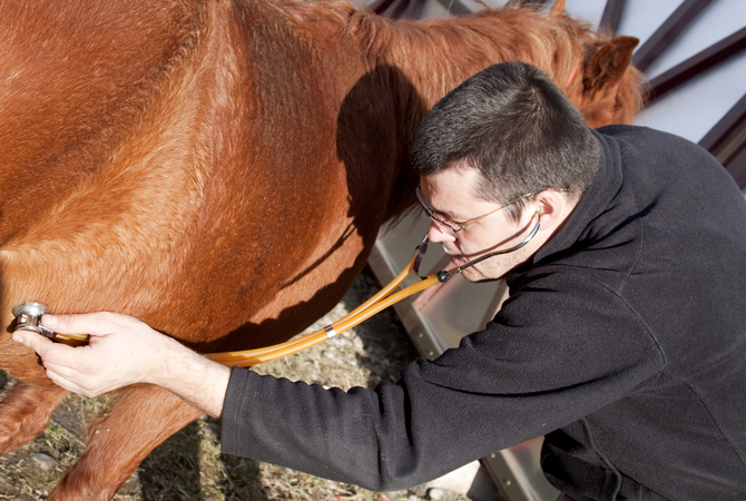
Image from azequine.com
Equine gastric ulcer syndrome (EGUS) is common in horses. Ulcers can be found in the terminal esophagus, stomach (nonglandular and glandular regions) and the proximal duodenum. Prevalence estimates range from 25 percent to 50 percent in foals and 60 percent to 90 percent in adult horses, depending on age, performance, and evaluated populations. A presumptive diagnosis of EGUS relies on recognizing clinical signs and response to treatment, while a definitive diagnosis is based on endoscopic examination.
The equine stomach is divided into two distinct anatomic regions, the esophageal or nonglandular region and the glandular region. Gastric ulcers are commonly located in the nonglandular region of the stomach adjacent to the Margo plicatus along the greater curvature and lesser curvature.
Gastric ulcers in the glandular region are located near the pyloric opening of the stomach and frequently associated with administration of nonsteroidal anti-inflammatory drugs (NSAIDs). The horse stomach’s parietal cells continuously secrete hydrochloric acid, and secretion occurs in the absence of feed material. Gastric emptying of a liquid meal occurs within 30 minutes, whereas complete gastric emptying of a roughage hay meal occurs in 24 hours.
EGUS results from a disequilibrium between mucosal aggressive factors (acids, pepsin and bile acids) and mucosal protective factors (mucus and bicarbonate). Since mucosal protective factors are more developed in the glandular mucosa of the equine stomach when compared to the nonglandular mucosa, different causative mechanisms lead to ulceration in these regions.
Ulcers in the nonglandular mucosa are primarily caused by prolonged exposure to hydrochloric acid, pepsin or bile acids. Ulcers occurring in this region are similar to gastroesophageal reflux disease syndrome (GERDS) in humans, since this region lacks well-developed intrinsic mucosal protective factors, similar to the esophagus.
The severity of nonglandular ulcers is probably related to time length of acid exposure. The nonglandular mucosa near the Margo plicatus is constantly exposed to these aggressive factors and this region is where gastric ulcers are frequently found.
Ulcers in the glandular mucosa are primarily caused by disruption of blood flow and decreased mucus and bicarbonate secretion, which results in back diffusion of hydrogen ions and damage to the underlying submucosa. Prostaglandin inhibition may play a major role in the pathogenesis of gastric ulcers in the of the equine stomach’s glandular region.
Gastric ulceration in the nonglandular mucosa is directly related to the degree and length of gastric acid exposure. Several factors have been implicated in causing ulceration, including fasting, gastric acid clearance (gastric motility and emptying) and aggressive substances in gastric juice (acids [hydrochloric and short-chain fatty acids], pepsin and bile acids). In humans with GERDS, acid clearance time and consequent exposure of the esophageal mucosa to potentially injurious agents is inversely proportional to the rate of gastric esophageal and gastric motility. Delayed gastric emptying or decreased gastric motility can increase exposure of the nonglandular mucosa to gastric acid and other aggressive factors leading to ulceration.
The prevalence of EGUS is high in the performance horse and may be because of prolonged exposure of acids to the nonglandular mucosa and feeding schedules, which include periods of fasting. Fasting has been shown to cause ulcers in the nonglandular mucosa of horses and lower stomach pH.
In one study, horses fed hay continuously had higher median 24-hour gastric juice pH (3.1), when compared to horses that were fasted. Also, high-roughage diets stimulate production of bicarbonate rich saliva, which may buffer gastric acid.
Furthermore, exercise may inhibit gastric emptying, especially in racehorses that perform at near maximal levels. Decreased gastric and esophageal motility and delayed gastric emptying have been implicated in causing GERDS in humans during exercise and may lead to gastric ulceration in the performance horses, especially the racehorse.
Other organic acids in the stomach may act synergistically with hydrochloric acid to play a role in the pathogenesis of EGUS. Recently, short-chain fatty acids (SCFAs)-acetic, butyric, propionic and valeric acids-fermentation byproducts of carbohydrates, were found to induce acid injury to the gastroesophageal (nonglandular) mucosa of pigs and horses. These SCFAs are highly lipid-soluble and penetrate the nonglandular mucosa of the stomach at low gastric pH. They also lead to cell acidification, cell swelling, inflammation and ulceration. Thus, SCFAs may play a role in EGUS pathogenesis, especially in horses fed high-grain diets. In a previous report, SCFAs were found to be present in the stomach of horses fed alfalfa and brome grass hay and grain.4 Also, Scott et al, recently reported Helicobacter spp.-specific DNA in the nonglandular and glandular mucosa of the horse with EGUS using polymerase chain reaction (PCR). Thus, Helicobactor spp. may play a role in persistent or recurring EGUS; antibiotics may be indicated in treatment of refractory EGUS cases.
Diagnosing EGUS
The diagnosis of EGUS is based on the presence of clinical signs and confirmation with endoscopic examination. Clinical signs in horses include poor appetite or failure to consume a meal, dullness, attitude changes, decreased performance, reluctance to train, poor body condition, rough hair coat, weight loss, bruxism (grinding of teeth), ptyalism (excess salivation), excessive recumbency and low-grade colic. A presumptive diagnosis of EGUS can be made on these typical clinical signs and response to therapy.
A definitive diagnosis can only be made using a video or fiberoptic endoscope. The endoscope must be at least 200 cm long and approximately 10 to 13 mm in diameter. A longer endoscope (280 to 300 cm) is necessary to observe the duodenum in adult horses. A shorter endoscope (110 to 140 cm) of 10 mm diameter is sufficient to see the stomach of foals.
Treatment Protocol
Inhibiting gastric acid secretion is the mainstay of gastric ulcer treatment in horses and a number of agents have been used for treatment and prevention. Currently, GastroGard (omeprazole, Merial Limited, Atlanta, Ga.), a proton ion pump blocker, is the only FDA-approved agent for treating EGUS. However, many pharmacologic agents and their doses have been published in the literature.
A recent report in the literature documented that diet may be an important factor in cause and prevention of EGUS.4 In that study, mean gastric juice pH was significantly higher (P<0.01) five hours after feeding alfalfa hay and grain when compared to feeding bromegrass hay. Also, the number and severity of nonglandular mucosa ulcers were significantly lower in the horses fed the alfalfa hay and grain diet. The authors speculated the high calcium (14.1 mg/g) and protein (21 percent) concentrations in the alfalfa hay and grain diet acted as a dietary antacid to increase stomach pH. Thus, diets high in protein and calcium may have a protective effect on the non-glandular mucosa in horses up to five hours after feeding.
No Comments